

一个碱性磷酸酶功能研究的故事


独家抢先看
文/惠觅宙
1989年,我前往世界排名前16的多伦多大学攻读博士学位。早在1984年,我在北京协和医院的导师是池之盛教授,当时任内分泌科主任,也是中国第一本《糖尿病学》教材的作者。
我知道,多伦多大学附属的多伦多总医院班廷研究所(Banting Institute)曾发现胰岛素,这一成就获得了诺贝尔奖。这一事实激励我申请去那里攻读博士研究生。一年后,我转到多伦多西奈山医院,开展碱性磷酸酶功能方面的研究。
当时,碱性磷酸酶普遍被认为只是细胞分化的一个标志物——特别是成骨细胞(骨形成细胞)的标志。换句话说,一旦碱性磷酸酶表达出来,就意味着成骨细胞已经分化,也就是说细胞已经成熟并具备成骨功能。
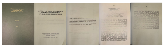
我当时的研究假说是:碱性磷酸酶不仅仅是一个细胞分化的标志物,它本身可能还具有生物学功能。这也成为我多伦多大学博士论文的核心题目——《寻找碱性磷酸酶的功能》(In Search of Functions of Alkaline Phosphatase)(图1)。
图1. 当时世界排名16的多伦多大学博士论文。
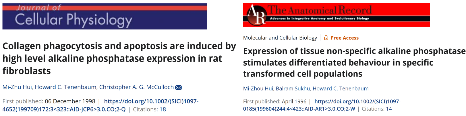
那时,我采用最早的分子生物学的方法,将碱性磷酸酶基因高表达在几种不同的细胞系中,包括成纤维样细胞、血管内皮细胞和肾小管上皮细胞(图2)。
图2. 碱性磷酸酶基因高表达在不同的细胞系明显影响细胞功能。
我所使用的细胞系是来自中国地仓鼠的多伦多CHO细胞系(图3)。有趣的是,这个多伦多CHO细胞系后来在生物制药行业中声名大噪,被广泛用于重组蛋白的表达与生产。
图3. 碱性磷酸酶基因高表达在CHO细胞系明显影响细胞形态和细胞增值。
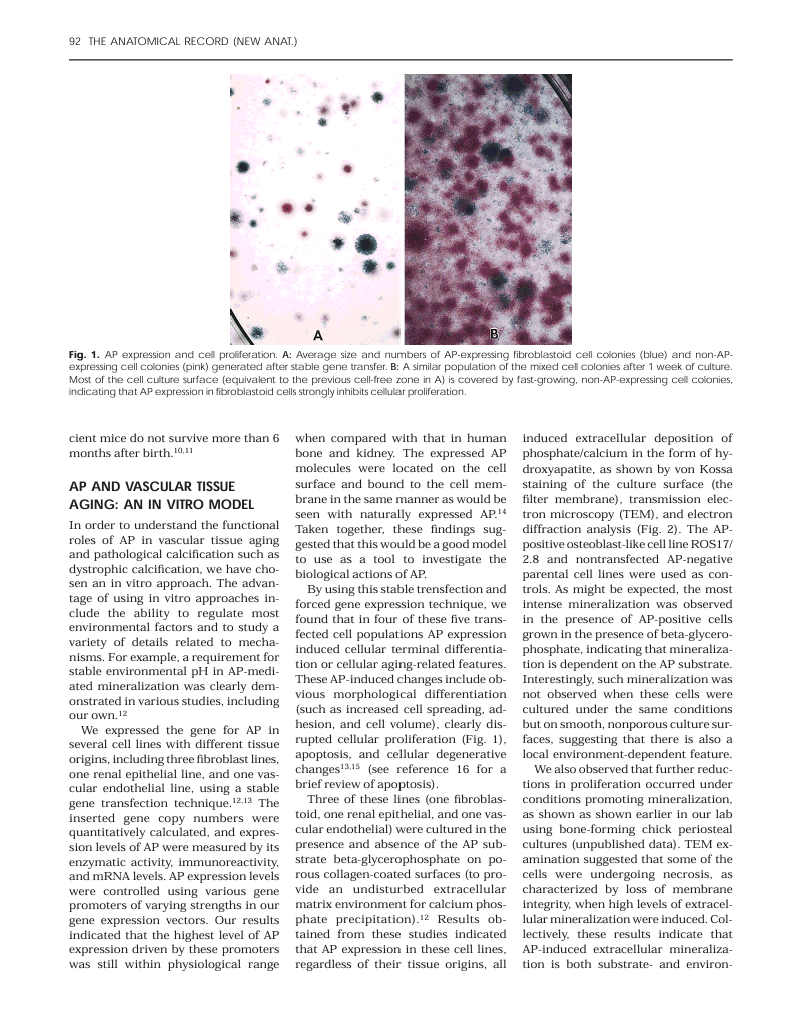
在这些细胞中表达碱性磷酸酶后,我观察到细胞形态和生长行为都发生了显著变化。换句话说,当碱性磷酸酶在细胞膜上高表达时,会诱导细胞形态的改变并抑制细胞生长。这是世界上首次发现碱性磷酸酶在细胞膜高表达时,在细胞水平上具有直接功能作用的研究。
我进一步发现,碱性磷酸酶的不同底物—如有机磷、ATP和LPS会产生不同的效应。当添加有机磷时,会产生磷沉积和细胞钙化作用。然而,即使不添加有机磷,也就是说仅有细胞自身产生的ATP、ADP和AMP时,碱性磷酸酶的细胞膜高表达仍然能抑制细胞生长并促进形态分化。
换句话说,我的研究结论是:碱性磷酸酶不仅是细胞成熟分化的标志物,还具有内在的生物学功能,可能是一种潜在的生物药物。
博士毕业后,我离开了碱性磷酸酶研究领域。直到2016年,我加入东北农业大学生命科学院,再次开始碱性磷酸酶功能研究。我发现,早在1997年,荷兰科学家Poelstra博士就已经发现碱性磷酸酶能够对内毒素(LPS)进行脱磷处理,从而降低其活性(1)。随后,荷兰一家制药公司(AM-Pharma)开发了重组碱性磷酸酶治疗脓毒血症相关内毒素血症的药物,并推进到临床三期试验(2)。
这些研究表明,依据底物的不同——无论是有机磷、ATP还是LPS—碱性磷酸酶都可以发挥不同的生物学功能。
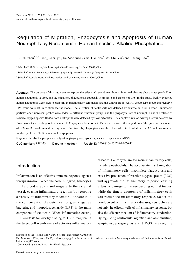
我进一步的研究证实,碱性磷酸酶确实在细胞水平上具有抗炎作用(图4),可望用于治疗多种炎症性疾病。
图4. 碱性磷酸酶在细胞水平抑制主要炎症细胞中性粒细胞的功能。
随后,通过查阅文献,我发现碱性磷酸酶的功能与两种著名的细胞外核苷酸酶—CD39和CD73—的功能基本一致。CD39⁺CD73⁺对T细胞的调节功能与碱性磷酸酶相同(3)。这些酶通过脱磷作用完成ATP–ADP–AMP–腺苷(AD)通路,减少促炎性的ATP、ADP和AMP,增加具有抗炎作用的腺苷(AD)(4)。
我现在希望通过基因治疗的方式,利用腺病毒载体高表达碱性磷酸酶,开发一种强效的低成本抗炎基因疗法,用于治疗炎症性疾病和炎症性创口。巧合的是计划中的动物和人体实验将采用与我在1990年代初期进行细胞基因转移研究相似的方法。
参考文献
Poelstra K, Bakker WW, Klok PA, Kamps JA, Hardonk MJ, Meijer DK. Dephosphorylation of endotoxin by alkaline phosphatase in vivo. Am J Pathol. 1997 Oct;151(4):1163-9.
Pickkers P, Angus DC, Bass K, Bellomo R, van den Berg E, Bernholz J, Bestle MH, Doi K, Doig CJ, Ferrer R, Francois B, Gammelager H, Pedersen UG, Hoste E, Iversen S, Joannidis M, Kellum JA, Liu K, Meersch M, Mehta R, Millington S, Murray PT, Nichol A, Ostermann M, Pettilä V, Solling C, Winkel M, Young PJ, Zarbock A; REVIVAL investigators. Phase-3 trial of recombinant human alkaline phosphatase for patients with sepsis-associated acute kidney injury (REVIVAL). Intensive Care Med. 2024 Jan;50(1):68-78.
Zhong EH, Ledderose C, De Andrade Mello P, Enjyoji K, Lunderberg JM, Junger W, Robson SC. Structural and functional characterization of engineered bifunctional fusion proteins of CD39 and CD73 ectonucleotidases. Am J Physiol Cell Physiol. 2021 Jan 1;320(1):C15-C29.
Antonioli L, Pacher P, Vizi ES, Haskó G. CD39 and CD73 in immunity and inflammation. Trends Mol Med. 2013 Jun;19(6):355-67.
The Story of Alkaline Phosphatase Function ResearchMizhou Hui
In 1989, I went to the University of Toronto—ranked among the world’s top 16 universities—to pursue my Ph.D. studies. Back in 1984, my mentor at Peking Union Medical College Hospital was Professor Chi Zhisheng, who was then the Director of the Department of Endocrinology and the author of China’s first textbook on diabetology.
I knew that the Banting Institute at Toronto General Hospital, affiliated with the University of Toronto, was where insulin was discovered—an achievement that won the Nobel Prize. This inspired me to apply for a Ph.D. position there. One year later, I transferred to Mount Sinai Hospital in Toronto to conduct research on the function of alkaline phosphatase.
At that time, alkaline phosphatase was widely regarded merely as a marker of cell differentiation—particularly for osteoblasts (bone-forming cells). In other words, once alkaline phosphatase was expressed, it indicated that the osteoblasts had differentiated, meaning they had matured and acquired bone-forming capability.
My research hypothesis at the time was that, beyond serving as a differentiation marker, alkaline phosphatase itself might have intrinsic biological functions. This became the central topic of my doctoral dissertation at the University of Toronto, titled “In Search of Functions of Alkaline Phosphatase” (Figure 1).
Figure 1. Ph.D. dissertation at the University of Toronto, which was ranked 16th in the world at the time.
At that time, I employed molecular biology techniques to overexpress the alkaline phosphatase gene in several different cell lines, including fibroblast-like cells, vascular endothelial cells, and renal tubular epithelial cells (Figure 2).
Figure 2. High expression of the alkaline phosphatase gene significantly affects cellular functions in different cell lines.
The fibroblast-like cells I used were the Chinese hamster ovary (CHO) cell line from Toronto (Figure 3. ). Interestingly, this Toronto CHO cell line later became widely known in the biopharmaceutical industry for its crucial role in recombinant protein expression and production.
Figure 3. High expression of the alkaline phosphatase gene in CHO cell lines significantly affects cell morphology and proliferation.
After expressing alkaline phosphatase in these cells, I observed notable changes in both cell morphology and growth behavior. In other words, when alkaline phosphatase was highly expressed on the cell surface, it induced morphological alterations and growth inhibition. This was the first discovery in the world demonstrating that high-level expression of alkaline phosphatase on the cell membrane had a direct functional effect at the cellular level.
I further found that different substrates of alkaline phosphatase—such as organic phosphate compounds, ATP, and LPS—produced distinct effects. When organic phosphate was added, phosphate deposition and cellular calcification occurred. However, even without organic phosphate, meaning only with endogenously produced ATP, ADP, and AMP, high membrane expression of alkaline phosphatase still inhibited cell growth and promoted morphological differentiation.
In other words, my research concluded that alkaline phosphatase is not only a marker of cell maturation and differentiation, but also possesses intrinsic biological functions, suggesting it could potentially serve as a biotherapeutic molecule.
After completing my Ph.D., I did not continue working in the field of alkaline phosphatase research. It was not until 2016, when I joined the Northeast Agricultural University Academy of Life Sciences, that I resumed my studies on alkaline phosphatase function. I found that in 1997, Dutch scientist Dr. Poelstra had already discovered that alkaline phosphatase can dephosphorylate endotoxin (LPS), thereby reducing its activity (1). Later, a Dutch pharmaceutical company (AM-Pharma) developed recombinant alkaline phosphatase therapy for sepsis-associated endotoxemia, which progressed to Phase III clinical trials (2).
These studies suggested that depending on the substrate—whether organic phosphate, ATP, or LPS—alkaline phosphatase exerts different biological functions.
My further research confirmed that alkaline phosphatase indeed has anti-inflammatory effects at the cellular level (Figure 4) and can potentially be used to treat various inflammatory diseases.
Figure 4. Alkaline phosphatase inhibits the function of neutrophils, the major inflammatory cells.
Later, through literature research, I discovered that the function of alkaline phosphatase is essentially identical to that of CD39 and CD73, two well-known ectonucleotidases. The regulatory effects of CD39⁺CD73⁺ on T cells are equivalent to the function of alkaline phosphatase (3). These enzymes complete the ATP–ADP–AMP–adenosine (AD) pathway through dephosphorylation—reducing the levels of the pro-inflammatory molecules ATP, ADP, and AMP, while increasing the anti-inflammatory molecule adenosine (AD) (4).
I now aim to apply gene therapy by using an adenoviral vector to achieve high expression of alkaline phosphatase, thereby developing a potent anti-inflammatory gene therapy for treating inflammatory diseases and inflammatory wounds. The planned studies in animals and humans will follow methods similar to those I used in the early 1990s for cellular gene transfer.
References
Poelstra K, Bakker WW, Klok PA, Kamps JA, Hardonk MJ, Meijer DK. Dephosphorylation of endotoxin by alkaline phosphatase in vivo. Am J Pathol. 1997 Oct;151(4):1163-9.
Pickkers P, Angus DC, Bass K, Bellomo R, van den Berg E, Bernholz J, Bestle MH, Doi K, Doig CJ, Ferrer R, Francois B, Gammelager H, Pedersen UG, Hoste E, Iversen S, Joannidis M, Kellum JA, Liu K, Meersch M, Mehta R, Millington S, Murray PT, Nichol A, Ostermann M, Pettilä V, Solling C, Winkel M, Young PJ, Zarbock A; REVIVAL investigators. Phase-3 trial of recombinant human alkaline phosphatase for patients with sepsis-associated acute kidney injury (REVIVAL). Intensive Care Med. 2024 Jan;50(1):68-78.
Zhong EH, Ledderose C, De Andrade Mello P, Enjyoji K, Lunderberg JM, Junger W, Robson SC. Structural and functional characterization of engineered bifunctional fusion proteins of CD39 and CD73 ectonucleotidases. Am J Physiol Cell Physiol. 2021 Jan 1;320(1):C15-C29.
Antonioli L, Pacher P, Vizi ES, Haskó G. CD39 and CD73 in immunity and inflammation. Trends Mol Med. 2013 Jun;19(6):355-67.


